Hechenyu Zha received her master’s degree from the School of Chemistry and Materials Science, University of Science and Technology of China, under the supervision of Prof. Xiang Shao. Her research mainly focused on the surface modification of metal oxide surface and its combination with two-dimensional materials
Xiang Shao received his Ph.D. degree in Physical Chemistry from Peking University. He is currently a Professor at the University of Science and Technology of China. His research interests cover the surface chemistry of metal alloys, metal oxides and their combinations with two-dimensional materials
Self-assembly films have demonstrated an efficient method to functionalize the surfaces of variously different materials. In this work, we preliminarily explored the modification effect of 10,12-pentacosadiynoic acid (PCDA) on the optical properties of monolayer molybdenum disulfide (MoS2) grown on a rutile titanium dioxide (r-TiO2) (110) single crystal surface. Atomic force microscopy (AFM) characterizations directly revealed that the PCDA molecules self-assemble into the same lamella structure as on pure MoS2, which can be further polymerized into conductive polydiacetylene (PDA) chains under ultraviolet light (UV) irradiation. Detailed photoluminescence (PL) measurements observed clearly increased luminescence of negative trions (A−) yet decreased total intensities for MoS2 upon adding the PCDA assembly, which is further enhanced after stimulating its polymerization. These results indicate that the PCDA assembly and its polymerization have different electron donability to MoS2, which hence provides a deepened understanding of the interfacial interactions within a multicomponent system. Our work also demonstrates the self-assembly of films as a versatile strategy to tune the electronic/optical properties of hybridized two-dimensional materials.
Graphical abstract
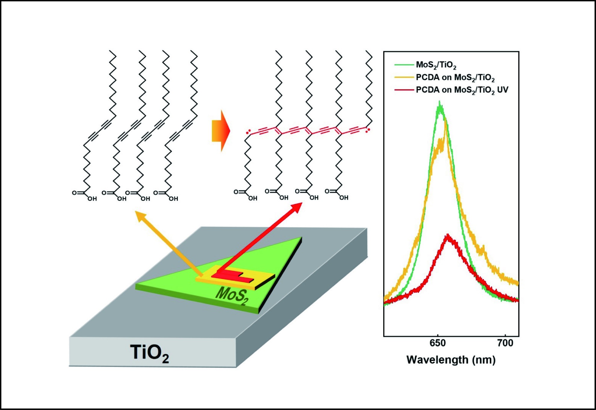
Self-assembly films of PCDA and its in situ photopolymerization donate different amount of electrons to the MoS2/TiO2 surface and thus modulate the photoluminescence of the latter.
Abstract
Self-assembly films have demonstrated an efficient method to functionalize the surfaces of variously different materials. In this work, we preliminarily explored the modification effect of 10,12-pentacosadiynoic acid (PCDA) on the optical properties of monolayer molybdenum disulfide (MoS2) grown on a rutile titanium dioxide (r-TiO2) (110) single crystal surface. Atomic force microscopy (AFM) characterizations directly revealed that the PCDA molecules self-assemble into the same lamella structure as on pure MoS2, which can be further polymerized into conductive polydiacetylene (PDA) chains under ultraviolet light (UV) irradiation. Detailed photoluminescence (PL) measurements observed clearly increased luminescence of negative trions (A−) yet decreased total intensities for MoS2 upon adding the PCDA assembly, which is further enhanced after stimulating its polymerization. These results indicate that the PCDA assembly and its polymerization have different electron donability to MoS2, which hence provides a deepened understanding of the interfacial interactions within a multicomponent system. Our work also demonstrates the self-assembly of films as a versatile strategy to tune the electronic/optical properties of hybridized two-dimensional materials.
Public Summary
Monolayer MoS2 was grown on rutile TiO2(110) single crystal through an ambient CVD method.
PCDA assembles into lamellae structure on the MoS2/TiO2 surface and polymerizes in situ under the UV irradiation.
The assembled and the further polymerized PCDA molecules donate electrons to the MoS2/TiO2 and tune the photoluminescence of the latter.
Recently, transition metal dichalcogenides (TMDs) have attracted immense research attention owing to their superior properties, including their layered structure, outstanding electrical conductivity and optical properties, and have been implemented in a wide range of applications, such as electronics, optics, sensors and heterogeneous catalysis[1–3]. In particular, when integrated with active oxide semiconductors, such as TiO2, the combinative system has been discovered with enhanced electro or photocatalytic performances[4–6] and thus has drawn intensive attention due to its expandable potential in electronics, photovoltaics, and photocatalysis. In the past several years, our group has developed an ambient-pressure chemical vapor deposition (CVD) method to grow high-quality single-layer MoS2 on differently terminated rutile TiO2 single crystals to successfully construct model hybridized MoS2/TiO2 systems with ideally controlled interfaces[7, 8]. Based on the series of characterizations, we revealed that the subtle change in the atomic structure of the interface may significantly influence the optical properties of the combinational system. Moreover, under most application conditions, the MoS2/TiO2 structure will be further integrated with other functional materials. In this case, the interactions between multiple interfaces must be carefully considered.
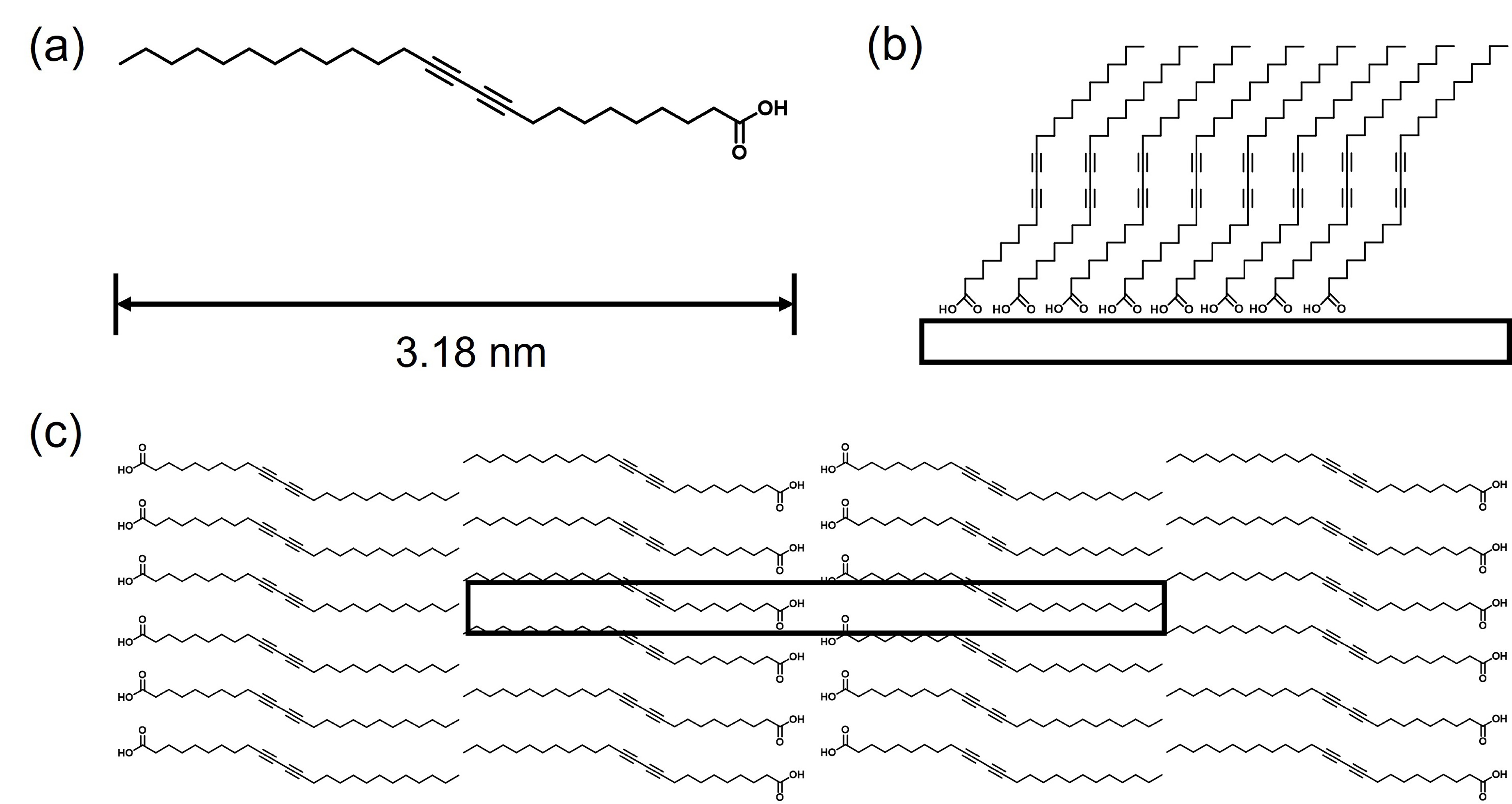
Organic self-assembly films (SAMs) have been demonstrated as an effective method to modify various surfaces in recent decades[9, 10]. In fact, SAMs are also frequently applied in solar cells or photovoltaic devices[11, 12]. Nevertheless, the detailed interactions between SAMs and the surfaces of functional semiconductors are not yet well understood, particularly for heterostructures comprising ultrathin two-dimensional materials (2DM). In this work, we investigated for the first time the assembly behavior of a prototypical photosensitive molecule, 10,12-pentacosadiynoic acid (PCDA, see Fig. 1), on the surface of the MoS2/TiO2 heterostructure. PCDA has been demonstrated to assemble into lamella structures on various surfaces[13, 14]. The aligned diacetylene groups can be further polymerized into linear conductive chains under ultraviolet (UV) light irradiation or pulse excitation and thus are considered promising candidates for molecular conductors[15]. Our atomic force microscopy (AFM) characterizations demonstrate that the PCDA molecules form the same assembly structure on the MoS2/TiO2 surface as on the pure MoS2 substrate and can also be triggered to polymerize upon UV irradiation. However, the molecular film and its reaction product have different effects on the photoluminescence of MoS2, which can be attributed to the different charge transfer conditions at the interface. These results may shed new light on the interface interactions between functional organic SAMs and hybridized semiconductive materials.
Figure
1.
(a) Molecular structure of 10,12-pentacosadiynoic acid. (b, c) The standing-up and flat-lying assembly structures of PCDA on oxide and other inert substrates, respectively. The rectangle in (c) marks the unit cell.
The monolayer MoS2/TiO2 substrate was prepared with the same recipe reported previously by our group[7, 8]. As the first step, the received rutile TiO2(110) single crystal (Hefei Kejing Materials Technology Co., Ltd.) was etched with HF solution, followed by annealing in air to obtain an atomically flat surface. Then, the as-prepared TiO2 substrate was loaded into the high-temperature zone of a two-zone tube furnace (Hefei Kejing Materials Technology Co., Ltd.). After that, 120 mg of sulfur powder (Shanghai Aladdin Biochemical Technology Co., Ltd., 99.95%) was weighed in a quartz boat and placed in the upstream low-temperature zone of the tube furnace, where the temperature was set at 200 °C. Meanwhile, 6 mg of molybdenum trioxide (MoO3, Shanghai Aladdin Biochemical Technology Co., Ltd., 99.95%) was weighed in another quartz boat and placed approximately 2 cm downstream against the TiO2 substrate. When everything was assembled, pure nitrogen gas was introduced at a gas rate of 300 sccm for 30 min to flush out the residual air and to ensure an inert gas environment during the reaction process. After that, the nitrogen flow rate was lowered to 100 sccm, and the high-temperature zone was gradually heated to ~750 °C at a speed of 15 °C/min to ensure that both the S and MoO3 precursors reached their sublimation temperature at roughly the same time. For the samples used in this study, the reaction was maintained for ~10 min. As a result, we could fabricate uniformly distributed monolayer MoS2 flakes as large as 1 µm for the side width while perfectly maintaining the atomic flatness of the surface, as will be presented below.
2.2
Preparation of the PCDA assembly films
All chemicals were used as received without further purification. The PCDA molecule (Shanghai Aladdin Biochemical Technology Co., Ltd., 97%) was dissolved in toluene (Sinopharm Chemical Reagent Co., Ltd., analytical purity) until the concentration reached 1.0×10⁻⁵–1.0×10⁻⁴ mol/L. The PCDA/toluene solutions were carefully stored in brown volumetric flasks and renewed after a few days. To prepare the SAMSs on different substrates, 10 µL of solution was drop cast onto the MoS2 or MoS2/TiO2 surface and kept under the cover of a beaker for a few minutes until the solvent completely evaporated. Commercial single-crystal MoS2 (Hefei Kejing Materials Technology Co., Ltd., 15 × 15 × 1 mm3) was cut into small squares (~5 mm × 5 mm width) before use. To prepare a clean surface, the top layers of the MoS2 crystal were either peeled off with tape or mechanically cleaved with a clean knife. In contrast, the MoS2/TiO2 substrate was directly used without further cleaning. All samples were characterized immediately after preparation. For the UV irradiation experiments, a UV lamp (TANK007, UV-AA01) with a wavelength centered at 365 nm and a power of 3 W was used, which was kept ~3 cm from the sample surface and illuminated for 5–10 min to stimulate the polymerization of PCDA. No obvious temperature rise of the sample was found.
2.3
Characterization methods
All microscopic measurements were acquired on a Digital Instruments NanoScope IIIa MultiMode™ system under ambient conditions. AFM images were acquired using a silicon cantilever probe (VTESPA-300, Bruker). Scanning tunneling microscopy (STM) images were acquired using a Pt/Ir tip prepared by mechanical cutting from a 0.25 mm platinum-iridium wire (Alfa Aesar). The STM experiments were only successfully performed for PCDA on the pure MoS2 surface, wherein a commercial MoS2 crystal (Hefei Kejing Materials Technology Co., Ltd.) was used as the substrate. Unfortunately, STM measurements of PCDA on MoS2/TiO2 did not succeed owing to the lack of sufficient conductivity. Photoluminescence (PL) measurements were carried out using a laser Raman spectrometer system from LabRamHR Evolution (JY, France). The incident laser beam has a wavelength of 532 nm and a power of 1.5 mW. The diameter of the focus is approximately 1 µm, which ideally fits the size of our as-grown MoS2. The integration time is normally set as 10 s.
3.
Results and discussion
3.1
Characterization of the as-prepared MoS2/TiO2 substrate
As stated in the experimental section, the MoS2/TiO2 substrate was fabricated through the ambient-pressure CVD recipe developed in our own group[8, 16]. In Fig. 2, we present the main characterization results of the MoS2/TiO2 sample. Fig. 2a displays the AFM image of the bare TiO2 surface before CVD synthesis of MoS2, which shows atomically flat terraces as wide as ~200 nm and separated by monoatomic steps (~0.34 nm for the height)[17]. After the CVD synthesis, the AFM image in Fig. 2b clearly shows a triangular-shaped monolayer MoS2 flake overlapping on the TiO2 surface while perfectly maintaining the surface flatness and cleanness. The inserted section profile across the MoS2 boundary directly reads the height of the MoS2 flake as ~0.71 nm, demonstrating its monolayer thickness[8]. Meanwhile, the step features of TiO2 underneath MoS2 can also be clearly observed, indicating that both materials have intimate contact. Fig. 2c shows a typical SEM image of the as-grown sample, manifesting the uniformly high quality of the fabricated MoS2, each having an average size of ~1µm.
Figure
2.
Characterization of the as-prepared MoS2/TiO2 substrate. (a) AFM image of an atomically flat TiO2(110) surface. The inset in the upper right corner shows the profile taken along the black arrow. (b) AFM image of a triangular MoS2 flake on the MoS2/TiO2 surface. The upper right inset shows the height profile measured across the MoS2 boundary along the black arrow. (c) Typical SEM image of the as-prepared MoS2/TiO2 surface. (d) Raman spectrum of MoS2 transferred from TiO2 onto a SiO2/Si substrate. (e, f) High-resolution XPS spectra of Mo 3d and S 2p on MoS2/TiO2.
In addition to this microscopic evidence, we also performed spectroscopy measurements to characterize the MoS2/TiO2 sample. The Raman spectrum was taken on the special sample prepared by transferring the as-grown MoS2 onto a SiO2/Si substrate. This is done because TiO2 has very strong Raman features overlapping with and hence covering those for the MoS2 adlayer. The spectrum in Fig. 2d clearly shows the typical E12g and A1g features of MoS2 positioning at 385.99 cm−1 and 405.58 cm−1, respectively. The wavenumber difference between the two peaks was found to be 19.59 cm−1 (< 20 cm−1), which also provides additional evidence for the monolayer thickness of the as-grown MoS2[18, 19]. Fig. 2e and 2f show the XPS spectra of the MoS2/TiO2 sample. The Mo high-resolution XPS spectrum clearly displays two distinct peaks positioned at 231.65 eV and 234.55 eV, which are consistent with the Mo4+ 3d5/2 and Mo4+ 3d3/2 components of MoS2, respectively, as reported in the literature[20, 21]. Correspondingly, the S 2p spectrum in Fig. 2f also confirms the existing oxidation state of sulfur as −2[22]. Quantitative elemental analysis reveals that the S/Mo ratio is approximately 2.1, indicating that a nearly stoichiometric MoS2 crystal has been fabricated on TiO2.
3.2
Self-assembly of PCDA on the MoS2 surface
The self-assembly of PCDA and similar derivatives on the MoS2 surface has been extensively investigated previously[14, 23]. Fig. 3 presents our own experimental data of PCDA on MoS2, providing a reference for the study of PDCA on MoS2/TiO2. As shown in Fig. 1c, on inert surfaces such as graphite and MoS2, the PCDA molecules tend to lie down and assemble into a lamellar structure wherein the diacetylene groups are aligned into chains. Here, our AFM scanning (Fig. 3a) also clearly recognizes the chain-like structures, which are representative of the PCDA molecular rows with an interval of approximately 7 nm. The ambient STM characterization in Fig. 3b directly resolves each PCDA molecule in the rows. The bright regions correspond to the alkyl chains, including the diacetylene groups, while the dark troughs correspond to the hydrogen-bonded carboxylic end groups. Detailed profile analyses in Fig. 3c and 3d reveal that the width of the PCDA rows is ~7.1 nm, in which the PCDA molecules are separated from each other with an interval of ~0.6 nm. All these features are perfectly consistent with the literature report as well as the molecular models, as shown in Fig. 1[14].
Figure
3.
(a) AFM and (b) the corresponding STM characterizations of the self-assembly film of PCDA formed on MoS2. Four PCDA molecular models are overlapped over the STM image. Note that although the film coverage slightly surpasses that of the monolayer, STM can only observe the structure of the bottom layer. (c, d) are the section profiles taken along the black and blue lines, respectively. The measured distances correspond to ten and double times the periodicities in these directions.
3.3
Self-assembly of PCDA on the MoS2/TiO2 surface
After the PCDA test experiments on pure MoS2, we then tried to explore its assembly behavior on the MoS2/TiO2 surface. Fig. 4a shows a typical AFM image of depositing 10 µL of the PCDA/toluene (1×10⁻⁴ mol/L) solution on a 5 mm×10 mm MoS2/TiO2 substrate. Interestingly, it was found that PCDA does not form a uniform film on the surface but instead forms distinct aggregates on the TiO2 and MoS2 regions. The rectangular-shaped tall islands correspond to the aggregates on the TiO2 surface, which are proposed to be multiple layers of tilting PCDA molecules, as schemed in Fig. 1b. Although this is the first study to observe such PCDA assembly on the rutile TiO2(110) surface, we do not discuss it in detail but only focus on the aggregates formed on the MoS2 region. Fig. 4b shows the topographic AFM image covering a MoS2 flake. It can be clearly seen that the deposited PCDA molecules have assembled into submonolayer films with a height of ~0.5 nm. In addition, the zoomed-in phase image in Fig. 4c displays an obvious parallel-line pattern with a periodic interval of ~6.9 nm (see the profile analysis in Fig. 4d). These characteristics demonstrate that the assembly structure of PCDA on the MoS2/TiO2 surface should be exactly the same as that on the pure MoS2 surface[24]. All the PCDA molecules lie on the surface and align their diacetylene groups together, as shown in the model in Fig. 1c. Such a flat-lying configuration of PCDA is not surprising considering that the MoS2/TiO2 surface is also barely inert and cannot directly bond to the carboxylic end group of PCDA. However, such a configuration ensures direct contact of the diacetylene group with the MoS2 surface and hence facilitates charge transfer between the two materials, as will be discussed later.
Figure
4.
AFM characterization of the assembly structure of PCDA on the MoS2/TiO2 surface. (a) Large-area topographic image showing the PCDA aggregates formed on the MoS2/TiO2 surface. (b) Zoomed-in topographic image focusing on the MoS2/TiO2 region covered by PCDA film. (c) Zoomed-in phase image showing the resolved assembly structure of PCDA on MoS2/TiO2. (d) Section profile taken along the white arrow in (c), which measures the periodicity of the PCDA assembly as ~6.9 nm.
3.4
UV-stimulated polymerization of PCDA molecules on MoS2/TiO2
The most attractive property of the PCDA assembly is its polymerization reaction under external excitations. Previous studies have demonstrated that PCDA on MoS2 can be stimulated to polymerize upon UV irradiation[14]. However, whether this reaction can take place on MoS2/TiO2 is still unknown. Therefore, we shed a 365 nm UV light onto the PCDA/MoS2/TiO2 sample for ~10 min. As shown in Fig. 5a, it can be clearly seen that some brighter lines developed while part of the original lower ones are still observed. Their differences become more apparent when examining the corresponding phase image, as shown in Fig. 5b. These brighter chains are attributed to polymerized PCDA (PDA) on the MoS2/TiO2 surface[15]. Their increased height (~0.14 nm, see Fig. 5c) relative to the unreacted PCDA molecules can be explained by the lifted poly-diacetylene groups, as shown by the inserted model in Fig. 5c. Aside from the polymerized PCDA films, we also noticed that the triangular MoS2 flake and the TiO2 surface remained unchanged, indicating that UV light had a limited influence on these surface structures.
Figure
5.
(a) AFM topography and (b) the corresponding phase images of the PCDA/MoS2/TiO2 surface after UV irradiation. The black and red ovals highlight the polymerized and unreacted PCDA assembly, respectively. (c) Section profiles taken along the black and red arrows in (a), showing the height difference between the reacted and unreacted PCDA film. Inserted are the simple schematics for the flat lying PCDA (lower) and the PDA polymer (upper), respectively.
The optical property is one of the most attractive properties of MoS2[25, 26]. To explore the effect of PCDA molecules on the photoluminescence of the MoS2/TiO2 combinational system, we performed PL measurements on a series of PCDA/MoS2/TiO2 samples. Fig. 6a–c presents mainly the A peaks of MoS2 at approximately 650 nm, which is the most prominent PL signal resulting from the cross-band annihilation of the A excitons in MoS2[16, 27]. Based on the peak-fitting analysis, all the PL spectra can be divided into two components. The short wavelength component (in blue) is always dominant and can be attributed to the neutral A exciton, while the long wavelength component (in yellow) can be attributed to the negatively charged A exciton (A−)[28]. Detailed analysis found that both the intensity of PL and the intensity ratio of A−/A (${I_{{{\rm{A}}^ - }}}/{I_{{{\rm{A}}^0}}} $) significantly vary for different samples. As summarized in Fig. 6d, the PL intensity is the largest for the bare MoS2/TiO2 sample but consecutively decreases on PCDA/MoS2/TiO2 and UV-illuminated PCDA/MoS2/TiO2 (termed PCDA/MoS2/TiO2-UV). In contrast, the ratio of ${I_{{{\rm{A}}^ - }}}/{I_{{{\rm{A}}^0}}}$ reverses the ordering, indicating that the A− luminescence becomes increasingly prominent upon adding PCDA onto the MoS2/TiO2 surface and stimulating its polymerization. Realizing that the A− exciton is a quasiparticle formed by combining a neutral exciton with a free electron[28], we propose that the number of free electrons in MoS2 actually varies for these different PCDA/MoS2/TiO2 samples. These free electrons may be donated from the PCDA assembly films. As revealed by the AFM measurements, the PCDA molecules take a flat-lying configuration on the MoS2 surface, which means that their electron-donating diacetylene groups can directly contact the MoS2 surface to efficiently transfer electrons into the latter. For polymerized PDCA, although the poly-diacetylene groups are slightly lifted away from the surface, their conductive wire structure is much more beneficial for transporting electrons than the single molecules and thus may donate more electrons into MoS2. Along with the added electrons, the formation probability of the A− exciton in MoS2 synchronically increases. Because the formation of each A− consumes an A0 exciton, this simultaneously leads to a decrease in the A0 exciton during the luminescence process. Moreover, it is well acknowledged that A− has a significantly lower luminescence probability than A0[28]. Consequently, the total PL intensity also decreases along with the attachment of the PCDA assembly and their in situ polymerization.
Figure
6.
PL spectra measured on the different samples: (a) bare MoS2/TiO2, (b) PCDA/MoS2/TiO2 and (c) PCDA/MoS2/TiO2 structure after UV irradiation. All the spectra are properly fit with two peaks, i.e., A0 and A−. (d) Plot of the PL intensities and the ${I_{{{\rm{A}}^ - }}}/{I_{{{\rm{A}}^0}}} $ ratios versus different samples.
In summary, we have explored the modulation effect of organic self-assembled films on the properties of a hybridized semiconductor system. The investigated PCDA molecules play an electron donative role to the MoS2/TiO2 compositional surface. Moreover, their assembly films can be further polymerized under proper excitations such as UV irradiation in this work. The polymerized PDA film possesses a highly conjugated skeleton and thus serves as a better electron donor to the surface. Their charge transfer to MoS2 hence significantly varies the luminescence property of the latter. In this regard, we once again witness the effective and volatile modification effect of the organic films. Our results also emphasize that during the real designs as well as the practical applications of the functional hybridized systems, the interfacial interactions must be carefully considered.
Acknowledgements:
This work was supported by the National Natural Science Foundation of China (22172152, 21872130) and the National Key Research and Development Program of China (2021YFA1502801).
Li Y, Duerloo K A, Wauson K, et al. Structural semiconductor-to-semimetal phase transition in two-dimensional materials induced by electrostatic gating. Nat. Commun.,2016, 7: 10671. doi: 10.1038/ncomms10671
[2]
Liu E, Fu Y, Wang Y, et al. Integrated digital inverters based on two-dimensional anisotropic ReS2 field-effect transistors. Nat. Commun.,2015, 6: 6991. doi: 10.1038/ncomms7991
[3]
Iqbal M W, Iqbal M Z, Khan M F, et al. Tailoring the electrical and photo-electrical properties of a WS2 field effect transistor by selective n-type chemical doping. RSC Adv.,2016, 6 (29): 24675–24682. doi: 10.1039/C6RA02390H
[4]
Chen B, Meng Y H, Sha J W, et al. Preparation of MoS2/TiO2 based nanocomposites for photocatalysis and rechargeable batteries: progress, challenges, and perspective. Nanoscale,2018, 10 (1): 34–68. doi: 10.1039/C7NR07366F
[5]
Li Y H, Cai C Z, Gu Y H, et al. Novel electronic properties of a new MoS2/TiO2 heterostructure and potential applications in solar cells and photocatalysis. Appl. Surf. Sci.,2017, 414: 34–40. doi: 10.1016/j.apsusc.2017.04.001
[6]
Tu W G, Li Y C, Kuai L B, et al. Construction of unique two-dimensional MoS2-TiO2 hybrid nanojunctions: MoS2 as a promising cost-effective cocatalyst toward improved photocatalytic reduction of CO2 to methanol. Nanoscale,2017, 9 (26): 9065–9070. doi: 10.1039/C7NR03238B
[7]
Xiang M M, Huang C X, Xing Y, et al. Modulated photoluminescence of single-layer MoS2 on various rutile TiO2 surfaces: implications for photocatalytic applications. ACS Appl. Nano Mater.,2022, 5 (5): 7609–7618. doi: 10.1021/acsanm.2c01754
[8]
Liu H H, Li Y, Xiang M M, et al. Single layered MoS2 directly grown on rutile TiO2(110) for enhanced interfacial charge transfer. ACS Nano,2019, 13 (5): 6083–6089. doi: 10.1021/acsnano.9b02608
[9]
Kagan C R, Mitzi D B, Dimitrakopoulos C D. Organic-inorganic hybrid materials as semiconducting channels in thin-film field-effect transistors. Science,1999, 286 (5441): 945–947. doi: 10.1126/science.286.5441.945
[10]
Jariwala D, Howell S L, Chen K S, et al. Hybrid, gate-tunable, van der Waals p-n heterojunctions from pentacene and MoS2. Nano Lett.,2016, 16 (1): 497–503. doi: 10.1021/acs.nanolett.5b04141
[11]
Yu S H, Lee Y, Jang S K, et al. Dye-sensitized MoS2 photodetector with enhanced spectral photoresponse. ACS Nano,2014, 8 (8): 8285–8291. doi: 10.1021/nn502715h
[12]
Yuan Y J, Lu H W, Ji Z G, et al. Enhanced visible-light-induced hydrogen evolution from water in a noble-metal-free system catalyzed by ZnTCPP-MoS2/TiO2 assembly. Chem. Eng. J.,2015, 275: 8–16. doi: 10.1016/j.cej.2015.04.015
[13]
Guo C, Xue J D, Cheng L X, et al. Two-dimensional self-assembly of diacetylenic acid derivatives and their light-induced polymerization on HOPG surfaces. Phys. Chem. Chem. Phys.,2017, 19 (24): 16213–16218. doi: 10.1039/C7CP02337E
[14]
Giridharagopal R, Kelly K F. Substrate-dependent properties of polydiacetylene nanowires on graphite and MoS2. ACS Nano,2008, 2 (8): 1571–1580. doi: 10.1021/nn800287x
[15]
Okawa Y, Akai-Kasaya M, Kuwahara Y, et al. Controlled chain polymerisation and chemical soldering for single-molecule electronics. Nanoscale,2012, 4 (10): 3013–3028. doi: 10.1039/c2nr30245d
[16]
Liu H H. Direct fabrication of 2D materials on oxide surface and investigation of the interface properties. Thesis. Hefei: University of Science and Technology of China, 2018. (in Chinese)
[17]
Yamamoto Y, Nakajima K, Ohsawa T, et al. Preparation of atomically smooth TiO2 single crystal surfaces and their photochemical property. Jpn. J. Appl. Phys.,2005, 44 (17): L511–L514. doi: 10.1143/JJAP.44.L511
[18]
Wu J X, Liu J P, Cui J, et al. Dual-phase MoS2 as a high-performance sodium-ion battery anode. J. Mater. Chem. A,2020, 8 (4): 2114–2122. doi: 10.1039/C9TA11913B
[19]
Lee C, Yan H, Brus L E, et al. Anomalous lattice vibrations of single-and few-layer MoS2. ACS Nano,2010, 4 (5): 2695–2700. doi: 10.1021/nn1003937
[20]
Ho Y T, Ma C H, Luong T T, et al. Layered MoS2 grown on c-sapphire by pulsed laser deposition. Phys. Status Solidi RRL,2015, 9 (3): 187–191. doi: 10.1002/pssr.201409561
[21]
Ahn C, Lee J, Kim H U, et al. Low-temperature synthesis of large-scale molybdenum disulfide thin films directly on a plastic substrate using plasma-enhanced chemical vapor deposition. Adv. Mater.,2015, 27 (35): 5223–5229. doi: 10.1002/adma.201501678
[22]
Hao S, Yang B C, Gao Y L. Unravelling merging behaviors and electrostatic properties of CVD-grown monolayer MoS2 domains. J. Chem. Phys.,2016, 145 (8): 084704. doi: 10.1063/1.4961509
[23]
Mandal S K, Okawa Y, Hasegawa T, et al. Rate-determining factors in the chain polymerization of molecules initiated by local single-molecule excitation. ACS Nano,2011, 5 (4): 2779–2786. doi: 10.1021/nn103231j
[24]
Verveniotis E, Okawa Y, Makarova M V, et al. Self-assembling diacetylene molecules on atomically flat insulators. Phys. Chem. Chem. Phys.,2016, 18: 31600–31605. doi: 10.1039/C6CP06749B
[25]
Splendiani A, Sun L, Zhang Y, et al. Emerging photoluminescence in monolayer MoS2. Nano Lett.,2010, 10 (4): 1271–1275. doi: 10.1021/nl903868w
[26]
Yuan Y J, Lu H W, Yu Z T, et al. Noble-metal-free molybdenum disulfide cocatalyst for photocatalytic hydrogen production. ChemSusChem,2015, 8 (24): 4113–4127. doi: 10.1002/cssc.201501203
[27]
van der Zande A M, Huang P Y, Chenet D A, et al. Grains and grain boundaries in highly crystalline monolayer molybdenum disulphide. Nat. Mater.,2013, 12 (6): 554–561. doi: 10.1038/nmat3633
[28]
Mak K F, He K, Lee C, et al. Tightly bound trions in monolayer MoS2. Nat. Mater.,2013, 12 (3): 207–211. doi: 10.1038/nmat3505
Figure
1.
(a) Molecular structure of 10,12-pentacosadiynoic acid. (b, c) The standing-up and flat-lying assembly structures of PCDA on oxide and other inert substrates, respectively. The rectangle in (c) marks the unit cell.
Figure
2.
Characterization of the as-prepared MoS2/TiO2 substrate. (a) AFM image of an atomically flat TiO2(110) surface. The inset in the upper right corner shows the profile taken along the black arrow. (b) AFM image of a triangular MoS2 flake on the MoS2/TiO2 surface. The upper right inset shows the height profile measured across the MoS2 boundary along the black arrow. (c) Typical SEM image of the as-prepared MoS2/TiO2 surface. (d) Raman spectrum of MoS2 transferred from TiO2 onto a SiO2/Si substrate. (e, f) High-resolution XPS spectra of Mo 3d and S 2p on MoS2/TiO2.
Figure
3.
(a) AFM and (b) the corresponding STM characterizations of the self-assembly film of PCDA formed on MoS2. Four PCDA molecular models are overlapped over the STM image. Note that although the film coverage slightly surpasses that of the monolayer, STM can only observe the structure of the bottom layer. (c, d) are the section profiles taken along the black and blue lines, respectively. The measured distances correspond to ten and double times the periodicities in these directions.
Figure
4.
AFM characterization of the assembly structure of PCDA on the MoS2/TiO2 surface. (a) Large-area topographic image showing the PCDA aggregates formed on the MoS2/TiO2 surface. (b) Zoomed-in topographic image focusing on the MoS2/TiO2 region covered by PCDA film. (c) Zoomed-in phase image showing the resolved assembly structure of PCDA on MoS2/TiO2. (d) Section profile taken along the white arrow in (c), which measures the periodicity of the PCDA assembly as ~6.9 nm.
Figure
5.
(a) AFM topography and (b) the corresponding phase images of the PCDA/MoS2/TiO2 surface after UV irradiation. The black and red ovals highlight the polymerized and unreacted PCDA assembly, respectively. (c) Section profiles taken along the black and red arrows in (a), showing the height difference between the reacted and unreacted PCDA film. Inserted are the simple schematics for the flat lying PCDA (lower) and the PDA polymer (upper), respectively.
Figure
6.
PL spectra measured on the different samples: (a) bare MoS2/TiO2, (b) PCDA/MoS2/TiO2 and (c) PCDA/MoS2/TiO2 structure after UV irradiation. All the spectra are properly fit with two peaks, i.e., A0 and A−. (d) Plot of the PL intensities and the ${I_{{{\rm{A}}^ - }}}/{I_{{{\rm{A}}^0}}} $ ratios versus different samples.
[1]
Li Y, Duerloo K A, Wauson K, et al. Structural semiconductor-to-semimetal phase transition in two-dimensional materials induced by electrostatic gating. Nat. Commun.,2016, 7: 10671. doi: 10.1038/ncomms10671
[2]
Liu E, Fu Y, Wang Y, et al. Integrated digital inverters based on two-dimensional anisotropic ReS2 field-effect transistors. Nat. Commun.,2015, 6: 6991. doi: 10.1038/ncomms7991
[3]
Iqbal M W, Iqbal M Z, Khan M F, et al. Tailoring the electrical and photo-electrical properties of a WS2 field effect transistor by selective n-type chemical doping. RSC Adv.,2016, 6 (29): 24675–24682. doi: 10.1039/C6RA02390H
[4]
Chen B, Meng Y H, Sha J W, et al. Preparation of MoS2/TiO2 based nanocomposites for photocatalysis and rechargeable batteries: progress, challenges, and perspective. Nanoscale,2018, 10 (1): 34–68. doi: 10.1039/C7NR07366F
[5]
Li Y H, Cai C Z, Gu Y H, et al. Novel electronic properties of a new MoS2/TiO2 heterostructure and potential applications in solar cells and photocatalysis. Appl. Surf. Sci.,2017, 414: 34–40. doi: 10.1016/j.apsusc.2017.04.001
[6]
Tu W G, Li Y C, Kuai L B, et al. Construction of unique two-dimensional MoS2-TiO2 hybrid nanojunctions: MoS2 as a promising cost-effective cocatalyst toward improved photocatalytic reduction of CO2 to methanol. Nanoscale,2017, 9 (26): 9065–9070. doi: 10.1039/C7NR03238B
[7]
Xiang M M, Huang C X, Xing Y, et al. Modulated photoluminescence of single-layer MoS2 on various rutile TiO2 surfaces: implications for photocatalytic applications. ACS Appl. Nano Mater.,2022, 5 (5): 7609–7618. doi: 10.1021/acsanm.2c01754
[8]
Liu H H, Li Y, Xiang M M, et al. Single layered MoS2 directly grown on rutile TiO2(110) for enhanced interfacial charge transfer. ACS Nano,2019, 13 (5): 6083–6089. doi: 10.1021/acsnano.9b02608
[9]
Kagan C R, Mitzi D B, Dimitrakopoulos C D. Organic-inorganic hybrid materials as semiconducting channels in thin-film field-effect transistors. Science,1999, 286 (5441): 945–947. doi: 10.1126/science.286.5441.945
[10]
Jariwala D, Howell S L, Chen K S, et al. Hybrid, gate-tunable, van der Waals p-n heterojunctions from pentacene and MoS2. Nano Lett.,2016, 16 (1): 497–503. doi: 10.1021/acs.nanolett.5b04141
[11]
Yu S H, Lee Y, Jang S K, et al. Dye-sensitized MoS2 photodetector with enhanced spectral photoresponse. ACS Nano,2014, 8 (8): 8285–8291. doi: 10.1021/nn502715h
[12]
Yuan Y J, Lu H W, Ji Z G, et al. Enhanced visible-light-induced hydrogen evolution from water in a noble-metal-free system catalyzed by ZnTCPP-MoS2/TiO2 assembly. Chem. Eng. J.,2015, 275: 8–16. doi: 10.1016/j.cej.2015.04.015
[13]
Guo C, Xue J D, Cheng L X, et al. Two-dimensional self-assembly of diacetylenic acid derivatives and their light-induced polymerization on HOPG surfaces. Phys. Chem. Chem. Phys.,2017, 19 (24): 16213–16218. doi: 10.1039/C7CP02337E
[14]
Giridharagopal R, Kelly K F. Substrate-dependent properties of polydiacetylene nanowires on graphite and MoS2. ACS Nano,2008, 2 (8): 1571–1580. doi: 10.1021/nn800287x
[15]
Okawa Y, Akai-Kasaya M, Kuwahara Y, et al. Controlled chain polymerisation and chemical soldering for single-molecule electronics. Nanoscale,2012, 4 (10): 3013–3028. doi: 10.1039/c2nr30245d
[16]
Liu H H. Direct fabrication of 2D materials on oxide surface and investigation of the interface properties. Thesis. Hefei: University of Science and Technology of China, 2018. (in Chinese)
[17]
Yamamoto Y, Nakajima K, Ohsawa T, et al. Preparation of atomically smooth TiO2 single crystal surfaces and their photochemical property. Jpn. J. Appl. Phys.,2005, 44 (17): L511–L514. doi: 10.1143/JJAP.44.L511
[18]
Wu J X, Liu J P, Cui J, et al. Dual-phase MoS2 as a high-performance sodium-ion battery anode. J. Mater. Chem. A,2020, 8 (4): 2114–2122. doi: 10.1039/C9TA11913B
[19]
Lee C, Yan H, Brus L E, et al. Anomalous lattice vibrations of single-and few-layer MoS2. ACS Nano,2010, 4 (5): 2695–2700. doi: 10.1021/nn1003937
[20]
Ho Y T, Ma C H, Luong T T, et al. Layered MoS2 grown on c-sapphire by pulsed laser deposition. Phys. Status Solidi RRL,2015, 9 (3): 187–191. doi: 10.1002/pssr.201409561
[21]
Ahn C, Lee J, Kim H U, et al. Low-temperature synthesis of large-scale molybdenum disulfide thin films directly on a plastic substrate using plasma-enhanced chemical vapor deposition. Adv. Mater.,2015, 27 (35): 5223–5229. doi: 10.1002/adma.201501678
[22]
Hao S, Yang B C, Gao Y L. Unravelling merging behaviors and electrostatic properties of CVD-grown monolayer MoS2 domains. J. Chem. Phys.,2016, 145 (8): 084704. doi: 10.1063/1.4961509
[23]
Mandal S K, Okawa Y, Hasegawa T, et al. Rate-determining factors in the chain polymerization of molecules initiated by local single-molecule excitation. ACS Nano,2011, 5 (4): 2779–2786. doi: 10.1021/nn103231j
[24]
Verveniotis E, Okawa Y, Makarova M V, et al. Self-assembling diacetylene molecules on atomically flat insulators. Phys. Chem. Chem. Phys.,2016, 18: 31600–31605. doi: 10.1039/C6CP06749B
[25]
Splendiani A, Sun L, Zhang Y, et al. Emerging photoluminescence in monolayer MoS2. Nano Lett.,2010, 10 (4): 1271–1275. doi: 10.1021/nl903868w
[26]
Yuan Y J, Lu H W, Yu Z T, et al. Noble-metal-free molybdenum disulfide cocatalyst for photocatalytic hydrogen production. ChemSusChem,2015, 8 (24): 4113–4127. doi: 10.1002/cssc.201501203
[27]
van der Zande A M, Huang P Y, Chenet D A, et al. Grains and grain boundaries in highly crystalline monolayer molybdenum disulphide. Nat. Mater.,2013, 12 (6): 554–561. doi: 10.1038/nmat3633
[28]
Mak K F, He K, Lee C, et al. Tightly bound trions in monolayer MoS2. Nat. Mater.,2013, 12 (3): 207–211. doi: 10.1038/nmat3505
Zha H C Y, Xing Y, Xiang M M, et al. Tuning the photoluminescence of MoS2/TiO2 by molecular self-assembly films. JUSTC, 2023, 53(12): 1206 doi: 10.52396/JUSTC-2023-0078
Figure 1. (a) Molecular structure of 10,12-pentacosadiynoic acid. (b, c) The standing-up and flat-lying assembly structures of PCDA on oxide and other inert substrates, respectively. The rectangle in (c) marks the unit cell.
Figure 2. Characterization of the as-prepared MoS2/TiO2 substrate. (a) AFM image of an atomically flat TiO2(110) surface. The inset in the upper right corner shows the profile taken along the black arrow. (b) AFM image of a triangular MoS2 flake on the MoS2/TiO2 surface. The upper right inset shows the height profile measured across the MoS2 boundary along the black arrow. (c) Typical SEM image of the as-prepared MoS2/TiO2 surface. (d) Raman spectrum of MoS2 transferred from TiO2 onto a SiO2/Si substrate. (e, f) High-resolution XPS spectra of Mo 3d and S 2p on MoS2/TiO2.
Figure 3. (a) AFM and (b) the corresponding STM characterizations of the self-assembly film of PCDA formed on MoS2. Four PCDA molecular models are overlapped over the STM image. Note that although the film coverage slightly surpasses that of the monolayer, STM can only observe the structure of the bottom layer. (c, d) are the section profiles taken along the black and blue lines, respectively. The measured distances correspond to ten and double times the periodicities in these directions.
Figure 4. AFM characterization of the assembly structure of PCDA on the MoS2/TiO2 surface. (a) Large-area topographic image showing the PCDA aggregates formed on the MoS2/TiO2 surface. (b) Zoomed-in topographic image focusing on the MoS2/TiO2 region covered by PCDA film. (c) Zoomed-in phase image showing the resolved assembly structure of PCDA on MoS2/TiO2. (d) Section profile taken along the white arrow in (c), which measures the periodicity of the PCDA assembly as ~6.9 nm.
Figure 5. (a) AFM topography and (b) the corresponding phase images of the PCDA/MoS2/TiO2 surface after UV irradiation. The black and red ovals highlight the polymerized and unreacted PCDA assembly, respectively. (c) Section profiles taken along the black and red arrows in (a), showing the height difference between the reacted and unreacted PCDA film. Inserted are the simple schematics for the flat lying PCDA (lower) and the PDA polymer (upper), respectively.
Figure 6. PL spectra measured on the different samples: (a) bare MoS2/TiO2, (b) PCDA/MoS2/TiO2 and (c) PCDA/MoS2/TiO2 structure after UV irradiation. All the spectra are properly fit with two peaks, i.e., A0 and A−. (d) Plot of the PL intensities and the ${I_{{{\rm{A}}^ - }}}/{I_{{{\rm{A}}^0}}} $ ratios versus different samples.