
| [1] |
Hospital A, Goñi J R, Orozco M, et al. Molecular dynamics simulations: advances and applications. Advances and Applications in Bioinformatics and Chemistry, 2015, 8: 37–47. doi: 10.2147/AABC.S70333
|
| [2] |
Hollingsworth S A, Dror R O. Molecular dynamics simulation for all. Neuron, 2018, 99 (6): 1129–1143. doi: 10.1016/j.neuron.2018.08.011
|
| [3] |
Van Der Spoel D, Lindahl E, Hess B, et al. GROMACS: fast, flexible, and free. Journal of Computational Chemistry, 2005, 26 (16): 1701–1718. doi: 10.1002/jcc.20291
|
| [4] |
Case D A, Cheatham III T E, Darden T, et al. The Amber biomolecular simulation programs. Journal of Computational Chemistry, 2005, 26 (16): 1668–1688. doi: 10.1002/jcc.20290
|
| [5] |
Phillips J C, Hardy D J, Maia J D C, et al. Scalable molecular dynamics on CPU and GPU architectures with NAMD. The Journal of Chemical Physics, 2020, 153 (4): 044130. doi: 10.1063/5.0014475
|
| [6] |
Brooks B R, Brooks III C L, Mackerell Jr A D, et al. CHARMM: the biomolecular simulation program. Journal of Computational Chemistry, 2009, 30 (10): 1545–1614. doi: 10.1002/jcc.21287
|
| [7] |
Berman H, Henrick K, Nakamura H. Announcing the worldwide Protein Data Bank. Nature Structural & Molecular Biology, 2003, 10 (12): 980. doi: 10.1038/nsb1203-980
|
| [8] |
wwPDB consortium. Protein Data Bank: the single global archive for 3D macromolecular structure data. Nucleic Acids Research, 2019, 47 (D1): D520–D528. doi: 10.1093/nar/gky949
|
| [9] |
Callaway J, Cummings M, Deroski B, et al. Protein Data Bank contents guide: Atomic coordinate entry format description. Upton: Brookhaven National Laboratory, 1996 .
|
| [10] |
Westbrook J D, Fitzgerald P M D. The PDB Format, mmCIF Formats, and Other Data Formats. In: Bourne P E, Weissig H, editors. Structural Bioinformatics. Hoboken: John Wiley & Sons, Inc. , 2003 .
|
| [11] |
Zhao G, Perilla J R, Yufenyuy E L, et al. Mature HIV-1 capsid structure by cryo-electron microscopy and all-atom molecular dynamics. Nature, 2013, 497 (7451): 643–646. doi: 10.1038/nature12162
|
| [12] |
Khalid S, Brandner A F, Juraschko N, et al. Computational microbiology of bacteria: Advancements in molecular dynamics simulations. Structure, 2023, 31 (11): 1320–1327. doi: 10.1016/j.str.2023.09.012
|
| [13] |
Chua E Y D, Mendez J H, Rapp M, et al. Better, faster, cheaper: recent advances in cryo–electron microscopy. Annual Review of Biochemistry, 2022, 91: 1–32. doi: 10.1146/annurev-biochem-032620-110705
|
| [14] |
Fitzgerald P M D, Berman H, Bourne P, et al. The mmCIF dictionary: community review and final approval. Acta Crystallographica Section A, 1996, 52: C575. doi: 10.1107/S0108767396076593
|
| [15] |
Hall S R, Allen F H, Brown I D. The crystallographic information file (CIF): a new standard archive file for crystallography. Acta Crystallographica Section A, 1991, 47(6): 655–685. doi: 10.1107/S010876739101067X
|
| [16] |
Berman H M, Kleywegt G J, Nakamura H, et al. The Protein Data Bank archive as an open data resource. Journal of Computer-Aided Molecular Design, 2014, 28 (10): 1009–1014. doi: 10.1007/s10822-014-9770-y
|
| [17] |
van Ginkel G, Pravda L, Dana J M, et al. PDBeCIF: an open-source mmCIF/CIF parsing and processing package. BMC Bioinformatics, 2021, 22 (1): 383. doi: 10.1186/s12859-021-04271-9
|
| [18] |
Weaver L H, Matthews B W. Structure of bacteriophage T4 lysozyme refined at 1.7 Å resolution. Journal of Molecular Biology, 1987, 193 (1): 189–199. doi: 10.1016/0022-2836(87)90636-X
|
| [19] |
Mosalaganti S, Obarska-Kosinska A, Siggel M, et al. AI-based structure prediction empowers integrative structural analysis of human nuclear pores. Science, 2022, 376 (6598): eabm9506. doi: 10.1126/science.abm9506
|
| [20] |
Case D A, Aktulga H M, Belfon K A A, et al. Amber 2021. San Francisco: University of California, 2021 .
|
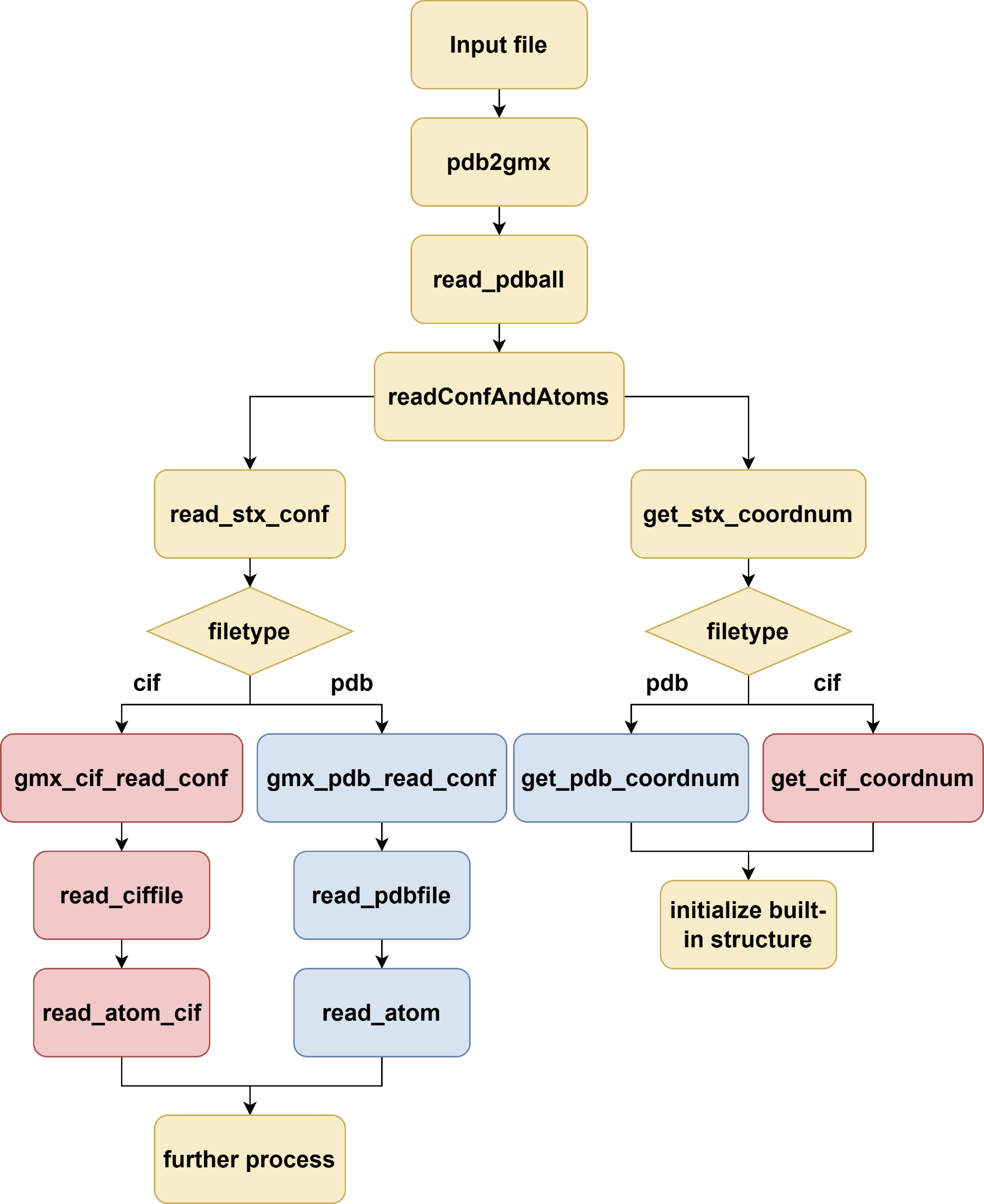
Figure 1. Flowchart of how GROMACS supports PDB and CIF files. Yellow: the same functions in processing PDB and CIF, red: PDB functions, and blue: CIF functions.

Figure 2. The systems used to test the modified GROMACS. (a) The bacteriophage T4 lysozyme (2LZM). (b) The dilated human nuclear pore complex (7R5J). Since the whole complex has C8 symmetry, only one-eighth of the structure is included in the CIF file.
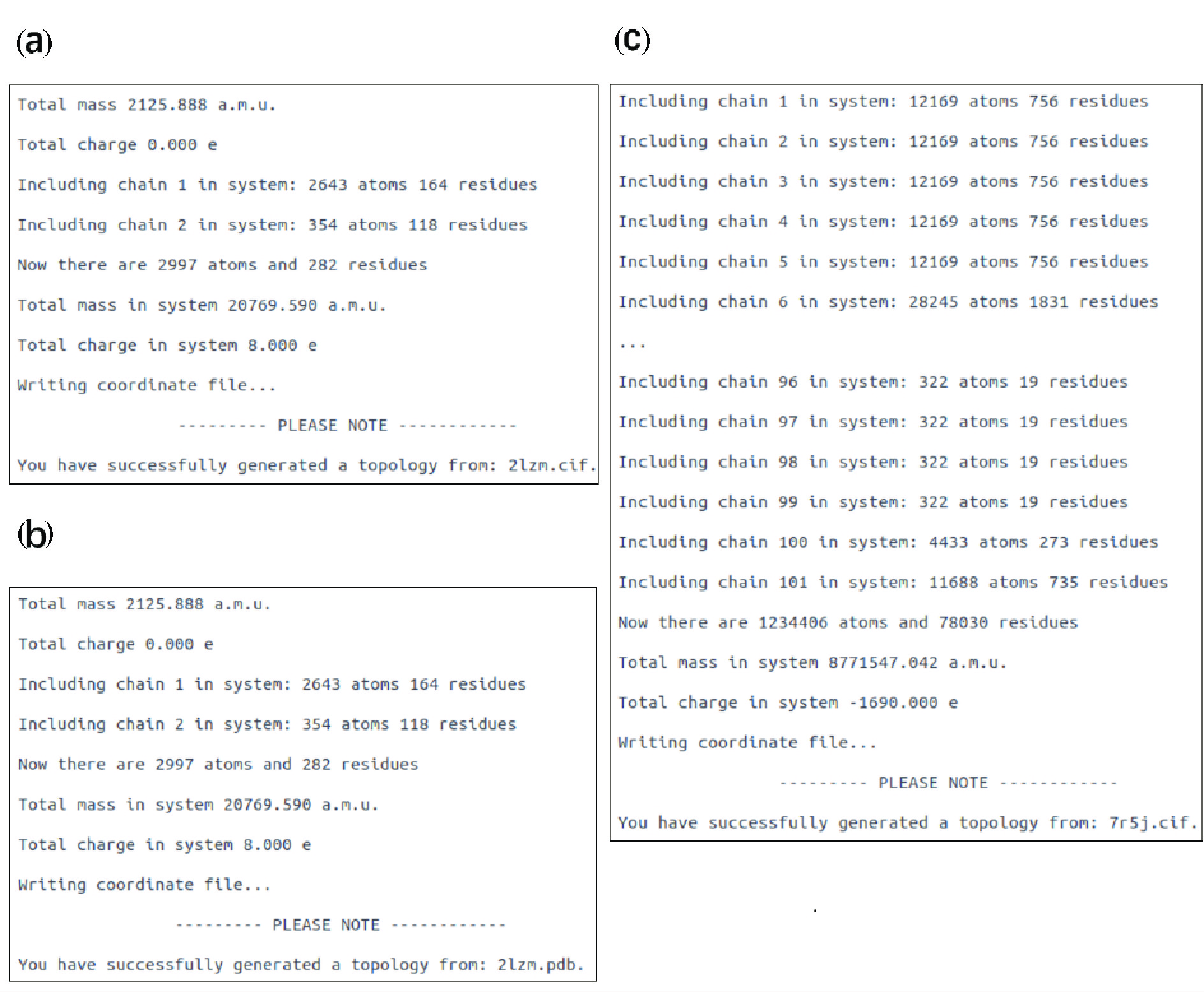
Figure 3. Screen output from the modified GROMACS. (a) Output when generating topology files from 2LZM.cif. (b) Output when generating topology files from 2LZM.pdb. (c) Output when generating topology files from 7R5J.cif.
| [1] |
Hospital A, Goñi J R, Orozco M, et al. Molecular dynamics simulations: advances and applications. Advances and Applications in Bioinformatics and Chemistry, 2015, 8: 37–47. doi: 10.2147/AABC.S70333
|
| [2] |
Hollingsworth S A, Dror R O. Molecular dynamics simulation for all. Neuron, 2018, 99 (6): 1129–1143. doi: 10.1016/j.neuron.2018.08.011
|
| [3] |
Van Der Spoel D, Lindahl E, Hess B, et al. GROMACS: fast, flexible, and free. Journal of Computational Chemistry, 2005, 26 (16): 1701–1718. doi: 10.1002/jcc.20291
|
| [4] |
Case D A, Cheatham III T E, Darden T, et al. The Amber biomolecular simulation programs. Journal of Computational Chemistry, 2005, 26 (16): 1668–1688. doi: 10.1002/jcc.20290
|
| [5] |
Phillips J C, Hardy D J, Maia J D C, et al. Scalable molecular dynamics on CPU and GPU architectures with NAMD. The Journal of Chemical Physics, 2020, 153 (4): 044130. doi: 10.1063/5.0014475
|
| [6] |
Brooks B R, Brooks III C L, Mackerell Jr A D, et al. CHARMM: the biomolecular simulation program. Journal of Computational Chemistry, 2009, 30 (10): 1545–1614. doi: 10.1002/jcc.21287
|
| [7] |
Berman H, Henrick K, Nakamura H. Announcing the worldwide Protein Data Bank. Nature Structural & Molecular Biology, 2003, 10 (12): 980. doi: 10.1038/nsb1203-980
|
| [8] |
wwPDB consortium. Protein Data Bank: the single global archive for 3D macromolecular structure data. Nucleic Acids Research, 2019, 47 (D1): D520–D528. doi: 10.1093/nar/gky949
|
| [9] |
Callaway J, Cummings M, Deroski B, et al. Protein Data Bank contents guide: Atomic coordinate entry format description. Upton: Brookhaven National Laboratory, 1996 .
|
| [10] |
Westbrook J D, Fitzgerald P M D. The PDB Format, mmCIF Formats, and Other Data Formats. In: Bourne P E, Weissig H, editors. Structural Bioinformatics. Hoboken: John Wiley & Sons, Inc. , 2003 .
|
| [11] |
Zhao G, Perilla J R, Yufenyuy E L, et al. Mature HIV-1 capsid structure by cryo-electron microscopy and all-atom molecular dynamics. Nature, 2013, 497 (7451): 643–646. doi: 10.1038/nature12162
|
| [12] |
Khalid S, Brandner A F, Juraschko N, et al. Computational microbiology of bacteria: Advancements in molecular dynamics simulations. Structure, 2023, 31 (11): 1320–1327. doi: 10.1016/j.str.2023.09.012
|
| [13] |
Chua E Y D, Mendez J H, Rapp M, et al. Better, faster, cheaper: recent advances in cryo–electron microscopy. Annual Review of Biochemistry, 2022, 91: 1–32. doi: 10.1146/annurev-biochem-032620-110705
|
| [14] |
Fitzgerald P M D, Berman H, Bourne P, et al. The mmCIF dictionary: community review and final approval. Acta Crystallographica Section A, 1996, 52: C575. doi: 10.1107/S0108767396076593
|
| [15] |
Hall S R, Allen F H, Brown I D. The crystallographic information file (CIF): a new standard archive file for crystallography. Acta Crystallographica Section A, 1991, 47(6): 655–685. doi: 10.1107/S010876739101067X
|
| [16] |
Berman H M, Kleywegt G J, Nakamura H, et al. The Protein Data Bank archive as an open data resource. Journal of Computer-Aided Molecular Design, 2014, 28 (10): 1009–1014. doi: 10.1007/s10822-014-9770-y
|
| [17] |
van Ginkel G, Pravda L, Dana J M, et al. PDBeCIF: an open-source mmCIF/CIF parsing and processing package. BMC Bioinformatics, 2021, 22 (1): 383. doi: 10.1186/s12859-021-04271-9
|
| [18] |
Weaver L H, Matthews B W. Structure of bacteriophage T4 lysozyme refined at 1.7 Å resolution. Journal of Molecular Biology, 1987, 193 (1): 189–199. doi: 10.1016/0022-2836(87)90636-X
|
| [19] |
Mosalaganti S, Obarska-Kosinska A, Siggel M, et al. AI-based structure prediction empowers integrative structural analysis of human nuclear pores. Science, 2022, 376 (6598): eabm9506. doi: 10.1126/science.abm9506
|
| [20] |
Case D A, Aktulga H M, Belfon K A A, et al. Amber 2021. San Francisco: University of California, 2021 .
|

ISSN 0253-2778
CN 34-1054/N
Copyright © Editorial Office of JUSTC, All Rights Reserved. 皖ICP备05002528号
Supported by:
Beijing Renhe Information Technology Co. Ltd


 DownLoad: