
| [1] |
Chauhan D, Vande Walle L, Lamkanfi M. Therapeutic modulation of inflammasome pathways. Immunological Reviews, 2020, 297 (1): 123–138. doi: 10.1111/imr.12908
|
| [2] |
McKee C M, Coll R C. NLRP3 inflammasome priming: A riddle wrapped in a mystery inside an enigma. Journal of Leukocyte Biology, 2020, 108 (3): 937–952. doi: 10.1002/JLB.3MR0720-513R
|
| [3] |
O’Connor W Jr, Harton J A, Zhu X, et al. Cutting edge: CIAS1/Cryopyrin/PYPAF1/NALP3/CATERPILLER 1.1 is an inducible inflammatory mediator with NF-kB suppressive properties. The Journal of Immunology, 2003, 171 (12): 6329–6333. doi: 10.4049/jimmunol.171.12.6329
|
| [4] |
Bauernfeind F G, Horvath G, Stutz A, et al. Cutting edge: NF-kB activating pattern recognition and cytokine receptors license NLRP3 inflammasome activation by regulating NLRP3 expression. The Journal of Immunology, 2009, 183 (2): 787–791. doi: 10.4049/jimmunol.0901363
|
| [5] |
He Y, Zeng M Y, Yang D, et al. NEK7 is an essential mediator of NLRP3 activation downstream of potassium efflux. Nature, 2016, 530 (7590): 354–357. doi: 10.1038/nature16959
|
| [6] |
Schmid-Burgk J L, Chauhan D, Schmidt T, et al. A genome-wide CRISPR (clustered regularly interspaced short palindromic repeats) screen identifies NEK7 as an essential component of NLRP3 inflammasome activation. Journal of Biological Chemistry, 2016, 291 (1): 103–109. doi: 10.1074/jbc.C115.700492
|
| [7] |
Broz P, Dixit V M. Inflammasomes: mechanism of assembly, regulation and signaling. Nature Reviews Immunology, 2016, 16 (7): 407–420. doi: 10.1038/nri.2016.58
|
| [8] |
Halle A, Hornung V, Petzold G C, et al. The NALP3 inflammasome is involved in the innate immune response to amyloid-β. Nature Immunology, 2008, 9 (8): 857–865. doi: 10.1038/ni.1636
|
| [9] |
Duewell P, Kono H, Rayner K J, et al. NLRP3 inflammasomes are required for atherogenesis and activated by cholesterol crystals. Nature, 2010, 464 (7293): 1357–1361. doi: 10.1038/nature08938
|
| [10] |
Stienstra R, Joosten L A, Koenen T, et al. The infammasome-mediated caspase-1 activation controls adipocyte diferentiation and insulin sensitivity. Cell Metabolism, 2010, 12 (6): 593–605. doi: 10.1016/j.cmet.2010.11.011
|
| [11] |
Stienstra R, van Diepen J A, Tack C J, et al. Infammasome is a central player in the induction of obesity and insulin resistance. Proceedings of the National Academy of Sciences of the United States of America, 2011, 108 (37): 15324–15329. doi: 10.1073/pnas.1100255108
|
| [12] |
Toldo S, Abbate A. The NLRP3 infammasome in acute myocardial infarction. Nature Reviews Cardiology, 2018, 15 (4): 203–214. doi: 10.1038/nrcardio.2017.161
|
| [13] |
Joosten L A, Netea M G, Mylona E, et al. Engagement of fatty acids with Toll-like receptor 2 drives interleukin-1β production via the ASC/caspase 1 pathway in monosodium urate monohydrate crystal-induced gouty arthritis. Arthritis& Rheumatism, 2010, 62 (11): 3237–3248. doi: 10.1002/art.27667
|
| [14] |
Hayward J A, Mathur A, Ngo C, et al. Cytosolic recognition of microbes and pathogens: Inflammasomes in action. Microbiology and Molecular Biology Reviews, 2018, 82 (4): e00015–e00018. doi: 10.1128/MMBR.00015-18
|
| [15] |
Rathinam V A K, Vanaja S K, Waggoner L, et al. TRIF licenses caspase-11-dependent NLRP3 inflammasome activation by gram-negative bacteria. Cell, 2012, 150 (3): 606–619. doi: 10.1016/j.cell.2012.07.007
|
| [16] |
Muñoz-Planillo R, Kuffa P, Martínez-Colón G. K+ efflux is the common trigger of NLRP3 inflammasome activation by bacterial toxins and particulate matter. Immunity, 2013, 38 (6): 1142–1153. doi: 10.1016/j.immuni.2013.05.016
|
| [17] |
Franchi L, Muñoz-Planillo R, Núñez G. Sensing and reacting to microbes through the inflammasomes. Nature Immunology, 2012, 13 (4): 325–332. doi: 10.1038/ni.2231
|
| [18] |
Negash A A, Olson R M, Griffin S, et al. Modulation of calcium signaling pathway by hepatitis C virus core protein stimulates NLRP3 inflammasome activation. PLoS Pathogens, 2019, 15 (2): e1007593. doi: 10.1371/journal.ppat.1007593
|
| [19] |
Chen I Y, Moriyama M, Chang M F, et al. Severe acute respiratory syndrome coronavirus viroporin 3a activates the NLRP3 inflammasome. Frontiers in Microbiology, 2019, 10: 50. doi: 10.3389/fmicb.2019.00050
|
| [20] |
Ichinohe T, Pang I K, Iwasaki A. Influenza virus activates inflammasomes via its intracellular M2 ion channel. Nature Immunology, 2010, 11 (5): 404–410. doi: 10.1038/ni.1861
|
| [21] |
Kanneganti T D, Kundu M, Green D R. Innate immune recognition of mtDNA—An undercover signal? Cell Metabolism, 2015, 21 (6): 793–794. doi: 10.1016/j.cmet.2015.05.019
|
| [22] |
Shimada K, Crother T R, Karlin J, et al. Oxidized mitochondrial DNA activates the NLRP3 inflammasome during apoptosis. Immunity, 2012, 36 (3): 401–414. doi: 10.1016/j.immuni.2012.01.009
|
| [23] |
Tschopp J, Schroder K. NLRP3 inflammasome activation: The convergence of multiple signaling pathways on ROS production. Nature Reviews Immunology, 2010, 10 (3): 210–215. doi: 10.1038/nri2725
|
| [24] |
Huang X P, Ludke A, Dhingra S, et al. Class II transactivator knockdown limits major histocompatibility complex II expression, diminishes immune rejection, and improves survival of allogeneicbone marrow stem cells in the infarcted heart. FASEB Journal, 2016, 30 (9): 3069–3082. doi: 10.1096/fj.201600331R
|
| [25] |
Halle A, Hornung V, Petzold G C, et al. The NALP3 inflammasome is involved in the innate immune response to amyloid-β. Nature Immunology, 2008, 9 (8): 857–865. doi: 10.1038/ni.1636
|
| [26] |
Duewell P, Kono H, Rayner K J, et al. NLRP3 inflammasomes are required for atherogenesis and activated by cholesterol crystals. Nature, 2010, 464 (7293): 1357–1361. doi: 10.1038/nature08938
|
| [27] |
Shi F S, Yang L F, Kouadir M, et al. The NALP3 inflammasome is involved in neurotoxic prion peptide-induced microglial activation. Journal of Neuroinflammation, 2012, 9 (1): 73. doi: 10.1186/1742-2094-9-73
|
| [28] |
Hafner-Bratkovič I, Benčina M, Fitzgerald K A, et al. NLRP3 inflammasome activation in macrophage cell lines by prion protein fibrils as the source of IL-1β and neuronal toxicity. Cellular and Molecular Life Sciences, 2012, 69 (24): 4215–4228. doi: 10.1007/s00018-012-1140-0
|
| [29] |
Shi F S, Yang Y, Kouadir M, et al. Inhibition of phagocytosis and lysosomal acidification suppresses neurotoxic prion peptide-induced NALP3 inflammasome activation in BV2 microglia. Journal of Neuroimmunology, 2013, 260 (1/2): 121–125. doi: 10.1016/j.jneuroim.2013.04.016
|
| [30] |
Dostert C, Guarda G, Romero J F, et al. Malarial hemozoin is a Nalp3 inflammasome activating danger signal. PLoS One, 2009, 4 (8): e6510. doi: 10.1371/journal.pone.0006510
|
| [31] |
Nakahira K, Haspel J A, Rathinam V A K, et al. Autophagy proteins regulate innate immune responses by inhibiting the release of mitochondrial DNA mediated by the NALP3 inflammasome. Nature Immunology, 2011, 12 (3): 222–230. doi: 10.1038/ni.1980
|
| [32] |
Zhou R B, Yazdi A S, Menu P, et al. A role for mitochondria in NLRP3 inflammasome activation. Nature, 2011, 469 (7329): 221–225. doi: 10.1038/nature09663
|
| [33] |
Sanman L E, Qian Y, Eisele N A, et al. Disruption of glycolytic flux is a signal for inflammasome signaling and pyroptotic cell death. eLife, 2016, 5: e13663. doi: 10.7554/eLife.13663
|
| [34] |
Zhou R B, Tardivel A, Thorens B, et al. Thioredoxin-interacting protein links oxidative stress to inflammasome activation. Nature Immunology, 2010, 11 (2): 136–140. doi: 10.1038/ni.1831
|
| [35] |
Bauernfeind F, Bartok E, Rieger A, et al. Cutting edge: Reactive oxygen species inhibitors block priming, but not activation, of the NLRP3 inflammasome. The Journal of Immunology, 2011, 187 (2): 613–617. doi: 10.4049/jimmunol.1100613
|
| [36] |
Jabaut J, Ather J L, Taracanova A, et al. Mitochondria-targeted drugs enhance Nlrp3 inflammasome-dependent IL-1β secretion in association with alterations in cellular redox and energy status. Free Radical Biology and Medicine, 2013, 60: 233–245. doi: 10.1016/j.freeradbiomed.2013.01.025
|
| [37] |
Shimada K, Crother T R, Karlin J, et al. Oxidized mitochondrial DNA activates the NLRP3 inflammasome during apoptosis. Immunity, 2012, 36 (3): 401–414. doi: 10.1016/j.immuni.2012.01.009
|
| [38] |
Park S, Woo J H, Hwang I, et al. Defective mitochondrial fission augments NLRP3 inflammasome activation. Scientific Reports, 2015, 5: 15489. doi: 10.1038/srep15489
|
| [39] |
Rocha M, Apostolova N, Diaz-Rua R, et al. Mitochondria and T2D: Role of autophagy, ER stress, and inflammasome. Trends in Endocrinology & Metabolism, 2020, 31 (10): 725–741. doi: 10.1016/j.tem.2020.03.004
|
| [40] |
Zhong Z, Liang S, Sanchez-Lopez E, et al. New mitochondrial DNA synthesis enables NLRP3 inflammasome activation. Nature, 2018, 560 (7717): 198–203. doi: 10.1038/s41586-018-0372-z
|
| [41] |
Guntuku L, Gangasani J K, Thummuri D, et al. IITZ-01, a novel potent lysosomotropic autophagy inhibitor, has single-agent antitumor efficacy in triple-negative breast cancer in vitro and in vivo. Oncogene, 2019, 38 (4): 581–595. doi: 10.1038/s41388-018-0446-2
|

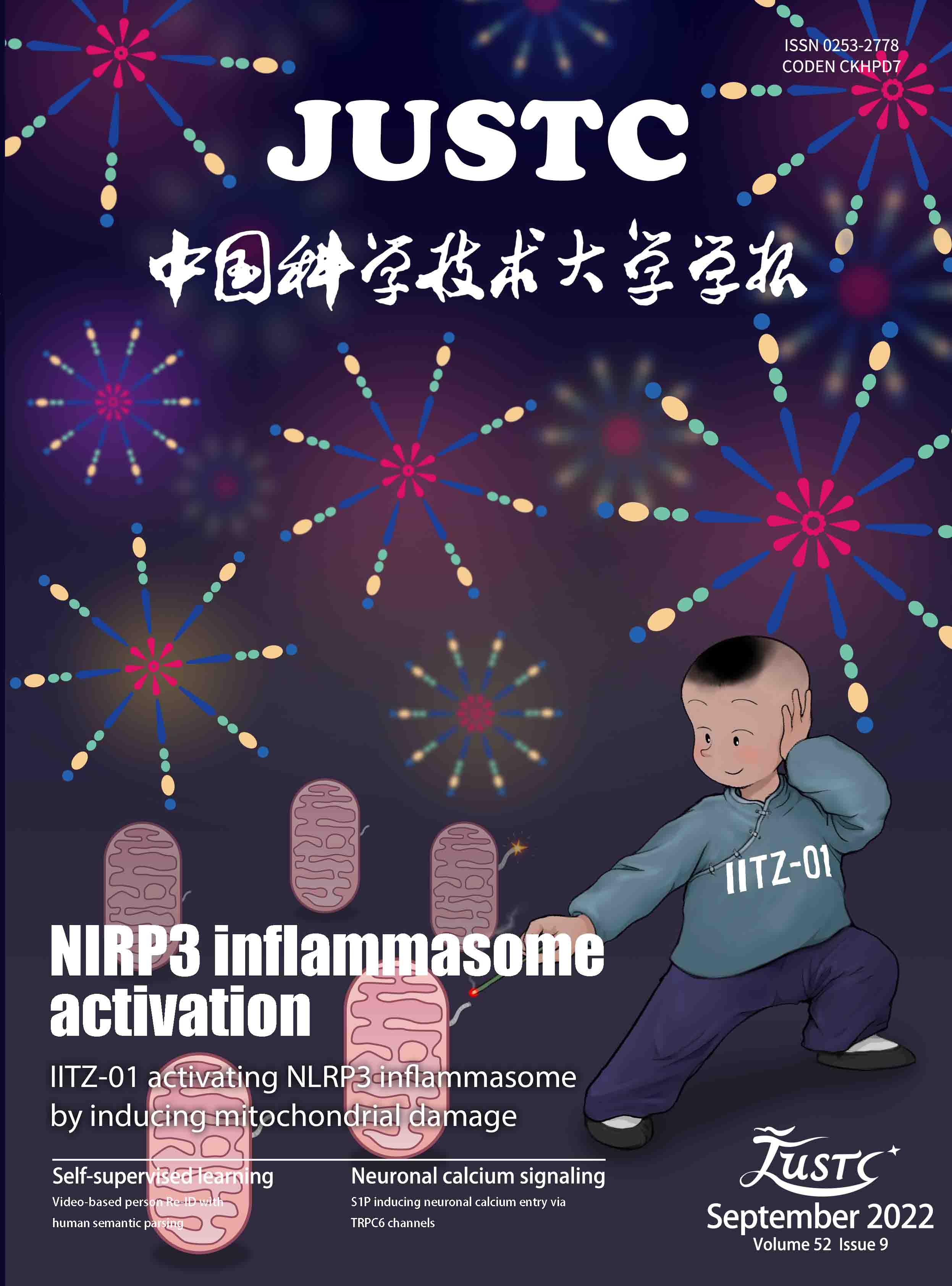
Figure 1. IITZ-01 induced caspase-1 activation and IL-1β production in BMDM cells. (a) ELISA of IL-1β in the supernatant from BMDM cells primed with LPS for 3 h and then stimulated with different doses of IITZ-01. (b, c) BMDM cells were primed with LPS for 3 h and then stimulated with IITZ-01 (2 μmol/L) for different times. (b) ELISA of IL-1β in the supernatant. (c) Western blot of IL-1β, cleaved caspase-1 in culture supernatant (SN) and the precursors of IL-1β (pro-IL-1β), pro-caspase-1, β–actin in cell lysate (input). Nigericin was used as a positive control. Data are means ± SEMs (n=6 or 5).

Figure
2.
IITZ-01 activated inflammasome in BMDM cells. (a, b) BMDM cells from WT or Casp1−/− mice were primed with LPS for 3 h and then stimulated with IITZ-01 (2 μmol/L) for different times. (a) ELISA of IL-1β in the supernatant. (b) Western blot of IL-1β and cleaved caspase-1 in SN and pro-IL-1β, pro-caspase-1, and β–actin in cell lysate. (c, d) BMDM cells from WT or Asc−/− mice were primed with LPS for 3 h and then stimulated with IITZ-01 (2 μmol/L) for different times. (c) ELISA of IL-1β in the supernatant. (d) Western blot of IL-1β and cleaved caspase-1 in SN and pro-IL-1β, pro-caspase-1, ASC, and β–actin in cell lysate. (e, f) BMDM cells from WT or Gsdmd−/− mice were primed with LPS for 3 h and then stimulated with IITZ-01 (2 μmol/L) for different times. (e) ELISA of IL-1β in the supernatant. (f) Western blot of IL-1β and cleaved caspase-1 in SN and pro-IL-1β, pro-caspase-1, GSDMD, and β–actin in cell lysate. Nigericin was used as a positive control. Data are means ± SEMs (n=5). **P

Figure
3.
IITZ-01 activated NLRP3 inflammasome in BMDM cells. (a, b) BMDM cells from WT or Nlrp3−/− mice were primed with LPS for 3 h and then stimulated with IITZ-01 (2 μmol/L) for different times. (a) ELISA of IL-1β in the supernatant. (b) Western blot of IL-1β and cleaved caspase-1 in SN and pro-IL-1β, pro-caspase-1, NLRP3, and β–actin in cell lysate. (c, d) BMDM cells were primed with LPS for 2 h, treated with different doses of CY-09 for 1 h, and then stimulated with IITZ-01 (2 μmol/L) for 3 h. (c) ELISA of IL-1β in the supernatant. (d) Western blot of IL-1β and cleaved caspase-1 in SN and pro-IL-1β, pro-caspase-1, and β–actin in cell lysate. (e, f) BMDM cells were primed with LPS for 2 h, treated with different doses of MCC950 for 1 h, and then stimulated with IITZ-01 (2 μmol/L) for 3 h. (e) ELISA of IL-1β in the supernatant. (f) Western blot of IL-1β and cleaved caspase-1 in SN and pro-IL-1β, pro-caspase-1, and β–actin in cell lysate. Nigericin was used as a positive control. Data are means ± SEMs (n=6, 5 or 5). *0.01

Figure 4. Inflammasome activation induced by IITZ-01 was independent of NLRC4, AIM2 and Pyrin. (a, b) BMDM cells from WT or Ipaf−/− mice were primed with LPS for 3 h and then stimulated with IITZ-01 (2 μmol/L) for different times. (a) ELISA of IL-1β in the supernatant. (b) Western blot of IL-1β and cleaved caspase-1 in SN and pro-IL-1β, pro-caspase-1, and β–actin in cell lysate. (c, d) BMDM cells from WT or Aim2−/− mice were primed with LPS for 3 h and then stimulated with IITZ-01 (2 μmol/L) for different times. (c) ELISA of IL-1β in the supernatant. (d) Western blot of IL-1β and cleaved caspase-1 in SN and pro-IL-1β, pro-caspase-1, and β–actin in cell lysate. (e, f) BMDM cells from WT or Pyrin−/− mice were primed with LPS for 3 h and then stimulated with IITZ-01 (2 μmol/L) for different times. (e) ELISA of IL-1β in the supernatant. (f) Western blot of IL-1β and cleaved caspase-1 in SN and pro-IL-1β, pro-caspase-1, and β–actin in cell lysate. Nigericin was used as a positive control. Data are means ± SEMs (n=4, 4 or 5). ns, not significant.

Figure 5. NLRP3 inflammasome activation induced by IITZ-01 was independent of direct binding and ion flow. (a) Western blot of NLRP3, NEK7, pro-caspase-1, ASC and β-actin in BMDM cells treated with DARTS assays. (b, c) ELISA of IL-1β in the supernatant from BMDM cells primed with LPS for 3 h, treated with different doses of KCl, (b) and then stimulated with IITZ-01 (2 μmol/L) for 3 h or (c) stimulated with Nigericin (5 μmol/L) for 15 min. (d) ICP of intracellular potassium in BMDM cells of Nlrp3−/− mice primed with LPS for 3 h and then stimulated with IITZ-01 (2 μmol/L) for 3 h or Nigericin (5 μmol/L) for 15 min. Data are means ± SEMs (n=6, 5 or 6).

Figure
6.
IITZ-01 activated NLRP3 inflammasome by inducing mitochondrial damage and mROS accumulation. (a) Immunofluorescence of mitochondrial morphology in BMDM cells from WT and Nlrp3−/− mice primed with LPS for 3 h and then stimulated with IITZ-01 (2 μmol/L) for 1 h or Nigericin (5 μmol/L) for 20 min. (b) Immunofluorescence of mROS in BMDM cells from WT and Nlrp3−/− mice primed with LPS for 3 h and then stimulated with IITZ-01 (2 μmol/L) for 3 h or Nigericin (5 μmol/L) for 20 min. (c) ELISA of IL-1β in the supernatant from BMDM cells primed with LPS for 2 h, treated with different doses of MnTBAP for 1 h, and then stimulated with IITZ-01 (2 μmol/L) for 5 h. (d) Western blot of IL-1β and cleaved caspase-1 in the SN of BMDM cells primed with LPS for 2 h, treated with different doses of MnTBAP for 1 h, and then stimulated with IITZ-01 (2 μmol/L) for 3 h or Nigericin (5 μmol/L) for 15 min. Western blot of pro-IL-1β, pro-caspase-1 and β-actin in the lysate of those cells. Data are means ± SEMs (n=5). ***P
| [1] |
Chauhan D, Vande Walle L, Lamkanfi M. Therapeutic modulation of inflammasome pathways. Immunological Reviews, 2020, 297 (1): 123–138. doi: 10.1111/imr.12908
|
| [2] |
McKee C M, Coll R C. NLRP3 inflammasome priming: A riddle wrapped in a mystery inside an enigma. Journal of Leukocyte Biology, 2020, 108 (3): 937–952. doi: 10.1002/JLB.3MR0720-513R
|
| [3] |
O’Connor W Jr, Harton J A, Zhu X, et al. Cutting edge: CIAS1/Cryopyrin/PYPAF1/NALP3/CATERPILLER 1.1 is an inducible inflammatory mediator with NF-kB suppressive properties. The Journal of Immunology, 2003, 171 (12): 6329–6333. doi: 10.4049/jimmunol.171.12.6329
|
| [4] |
Bauernfeind F G, Horvath G, Stutz A, et al. Cutting edge: NF-kB activating pattern recognition and cytokine receptors license NLRP3 inflammasome activation by regulating NLRP3 expression. The Journal of Immunology, 2009, 183 (2): 787–791. doi: 10.4049/jimmunol.0901363
|
| [5] |
He Y, Zeng M Y, Yang D, et al. NEK7 is an essential mediator of NLRP3 activation downstream of potassium efflux. Nature, 2016, 530 (7590): 354–357. doi: 10.1038/nature16959
|
| [6] |
Schmid-Burgk J L, Chauhan D, Schmidt T, et al. A genome-wide CRISPR (clustered regularly interspaced short palindromic repeats) screen identifies NEK7 as an essential component of NLRP3 inflammasome activation. Journal of Biological Chemistry, 2016, 291 (1): 103–109. doi: 10.1074/jbc.C115.700492
|
| [7] |
Broz P, Dixit V M. Inflammasomes: mechanism of assembly, regulation and signaling. Nature Reviews Immunology, 2016, 16 (7): 407–420. doi: 10.1038/nri.2016.58
|
| [8] |
Halle A, Hornung V, Petzold G C, et al. The NALP3 inflammasome is involved in the innate immune response to amyloid-β. Nature Immunology, 2008, 9 (8): 857–865. doi: 10.1038/ni.1636
|
| [9] |
Duewell P, Kono H, Rayner K J, et al. NLRP3 inflammasomes are required for atherogenesis and activated by cholesterol crystals. Nature, 2010, 464 (7293): 1357–1361. doi: 10.1038/nature08938
|
| [10] |
Stienstra R, Joosten L A, Koenen T, et al. The infammasome-mediated caspase-1 activation controls adipocyte diferentiation and insulin sensitivity. Cell Metabolism, 2010, 12 (6): 593–605. doi: 10.1016/j.cmet.2010.11.011
|
| [11] |
Stienstra R, van Diepen J A, Tack C J, et al. Infammasome is a central player in the induction of obesity and insulin resistance. Proceedings of the National Academy of Sciences of the United States of America, 2011, 108 (37): 15324–15329. doi: 10.1073/pnas.1100255108
|
| [12] |
Toldo S, Abbate A. The NLRP3 infammasome in acute myocardial infarction. Nature Reviews Cardiology, 2018, 15 (4): 203–214. doi: 10.1038/nrcardio.2017.161
|
| [13] |
Joosten L A, Netea M G, Mylona E, et al. Engagement of fatty acids with Toll-like receptor 2 drives interleukin-1β production via the ASC/caspase 1 pathway in monosodium urate monohydrate crystal-induced gouty arthritis. Arthritis& Rheumatism, 2010, 62 (11): 3237–3248. doi: 10.1002/art.27667
|
| [14] |
Hayward J A, Mathur A, Ngo C, et al. Cytosolic recognition of microbes and pathogens: Inflammasomes in action. Microbiology and Molecular Biology Reviews, 2018, 82 (4): e00015–e00018. doi: 10.1128/MMBR.00015-18
|
| [15] |
Rathinam V A K, Vanaja S K, Waggoner L, et al. TRIF licenses caspase-11-dependent NLRP3 inflammasome activation by gram-negative bacteria. Cell, 2012, 150 (3): 606–619. doi: 10.1016/j.cell.2012.07.007
|
| [16] |
Muñoz-Planillo R, Kuffa P, Martínez-Colón G. K+ efflux is the common trigger of NLRP3 inflammasome activation by bacterial toxins and particulate matter. Immunity, 2013, 38 (6): 1142–1153. doi: 10.1016/j.immuni.2013.05.016
|
| [17] |
Franchi L, Muñoz-Planillo R, Núñez G. Sensing and reacting to microbes through the inflammasomes. Nature Immunology, 2012, 13 (4): 325–332. doi: 10.1038/ni.2231
|
| [18] |
Negash A A, Olson R M, Griffin S, et al. Modulation of calcium signaling pathway by hepatitis C virus core protein stimulates NLRP3 inflammasome activation. PLoS Pathogens, 2019, 15 (2): e1007593. doi: 10.1371/journal.ppat.1007593
|
| [19] |
Chen I Y, Moriyama M, Chang M F, et al. Severe acute respiratory syndrome coronavirus viroporin 3a activates the NLRP3 inflammasome. Frontiers in Microbiology, 2019, 10: 50. doi: 10.3389/fmicb.2019.00050
|
| [20] |
Ichinohe T, Pang I K, Iwasaki A. Influenza virus activates inflammasomes via its intracellular M2 ion channel. Nature Immunology, 2010, 11 (5): 404–410. doi: 10.1038/ni.1861
|
| [21] |
Kanneganti T D, Kundu M, Green D R. Innate immune recognition of mtDNA—An undercover signal? Cell Metabolism, 2015, 21 (6): 793–794. doi: 10.1016/j.cmet.2015.05.019
|
| [22] |
Shimada K, Crother T R, Karlin J, et al. Oxidized mitochondrial DNA activates the NLRP3 inflammasome during apoptosis. Immunity, 2012, 36 (3): 401–414. doi: 10.1016/j.immuni.2012.01.009
|
| [23] |
Tschopp J, Schroder K. NLRP3 inflammasome activation: The convergence of multiple signaling pathways on ROS production. Nature Reviews Immunology, 2010, 10 (3): 210–215. doi: 10.1038/nri2725
|
| [24] |
Huang X P, Ludke A, Dhingra S, et al. Class II transactivator knockdown limits major histocompatibility complex II expression, diminishes immune rejection, and improves survival of allogeneicbone marrow stem cells in the infarcted heart. FASEB Journal, 2016, 30 (9): 3069–3082. doi: 10.1096/fj.201600331R
|
| [25] |
Halle A, Hornung V, Petzold G C, et al. The NALP3 inflammasome is involved in the innate immune response to amyloid-β. Nature Immunology, 2008, 9 (8): 857–865. doi: 10.1038/ni.1636
|
| [26] |
Duewell P, Kono H, Rayner K J, et al. NLRP3 inflammasomes are required for atherogenesis and activated by cholesterol crystals. Nature, 2010, 464 (7293): 1357–1361. doi: 10.1038/nature08938
|
| [27] |
Shi F S, Yang L F, Kouadir M, et al. The NALP3 inflammasome is involved in neurotoxic prion peptide-induced microglial activation. Journal of Neuroinflammation, 2012, 9 (1): 73. doi: 10.1186/1742-2094-9-73
|
| [28] |
Hafner-Bratkovič I, Benčina M, Fitzgerald K A, et al. NLRP3 inflammasome activation in macrophage cell lines by prion protein fibrils as the source of IL-1β and neuronal toxicity. Cellular and Molecular Life Sciences, 2012, 69 (24): 4215–4228. doi: 10.1007/s00018-012-1140-0
|
| [29] |
Shi F S, Yang Y, Kouadir M, et al. Inhibition of phagocytosis and lysosomal acidification suppresses neurotoxic prion peptide-induced NALP3 inflammasome activation in BV2 microglia. Journal of Neuroimmunology, 2013, 260 (1/2): 121–125. doi: 10.1016/j.jneuroim.2013.04.016
|
| [30] |
Dostert C, Guarda G, Romero J F, et al. Malarial hemozoin is a Nalp3 inflammasome activating danger signal. PLoS One, 2009, 4 (8): e6510. doi: 10.1371/journal.pone.0006510
|
| [31] |
Nakahira K, Haspel J A, Rathinam V A K, et al. Autophagy proteins regulate innate immune responses by inhibiting the release of mitochondrial DNA mediated by the NALP3 inflammasome. Nature Immunology, 2011, 12 (3): 222–230. doi: 10.1038/ni.1980
|
| [32] |
Zhou R B, Yazdi A S, Menu P, et al. A role for mitochondria in NLRP3 inflammasome activation. Nature, 2011, 469 (7329): 221–225. doi: 10.1038/nature09663
|
| [33] |
Sanman L E, Qian Y, Eisele N A, et al. Disruption of glycolytic flux is a signal for inflammasome signaling and pyroptotic cell death. eLife, 2016, 5: e13663. doi: 10.7554/eLife.13663
|
| [34] |
Zhou R B, Tardivel A, Thorens B, et al. Thioredoxin-interacting protein links oxidative stress to inflammasome activation. Nature Immunology, 2010, 11 (2): 136–140. doi: 10.1038/ni.1831
|
| [35] |
Bauernfeind F, Bartok E, Rieger A, et al. Cutting edge: Reactive oxygen species inhibitors block priming, but not activation, of the NLRP3 inflammasome. The Journal of Immunology, 2011, 187 (2): 613–617. doi: 10.4049/jimmunol.1100613
|
| [36] |
Jabaut J, Ather J L, Taracanova A, et al. Mitochondria-targeted drugs enhance Nlrp3 inflammasome-dependent IL-1β secretion in association with alterations in cellular redox and energy status. Free Radical Biology and Medicine, 2013, 60: 233–245. doi: 10.1016/j.freeradbiomed.2013.01.025
|
| [37] |
Shimada K, Crother T R, Karlin J, et al. Oxidized mitochondrial DNA activates the NLRP3 inflammasome during apoptosis. Immunity, 2012, 36 (3): 401–414. doi: 10.1016/j.immuni.2012.01.009
|
| [38] |
Park S, Woo J H, Hwang I, et al. Defective mitochondrial fission augments NLRP3 inflammasome activation. Scientific Reports, 2015, 5: 15489. doi: 10.1038/srep15489
|
| [39] |
Rocha M, Apostolova N, Diaz-Rua R, et al. Mitochondria and T2D: Role of autophagy, ER stress, and inflammasome. Trends in Endocrinology & Metabolism, 2020, 31 (10): 725–741. doi: 10.1016/j.tem.2020.03.004
|
| [40] |
Zhong Z, Liang S, Sanchez-Lopez E, et al. New mitochondrial DNA synthesis enables NLRP3 inflammasome activation. Nature, 2018, 560 (7717): 198–203. doi: 10.1038/s41586-018-0372-z
|
| [41] |
Guntuku L, Gangasani J K, Thummuri D, et al. IITZ-01, a novel potent lysosomotropic autophagy inhibitor, has single-agent antitumor efficacy in triple-negative breast cancer in vitro and in vivo. Oncogene, 2019, 38 (4): 581–595. doi: 10.1038/s41388-018-0446-2
|

ISSN 0253-2778
CN 34-1054/N
Copyright © Editorial Office of JUSTC, All Rights Reserved. 皖ICP备05002528号
Supported by:
Beijing Renhe Information Technology Co. Ltd


 DownLoad: